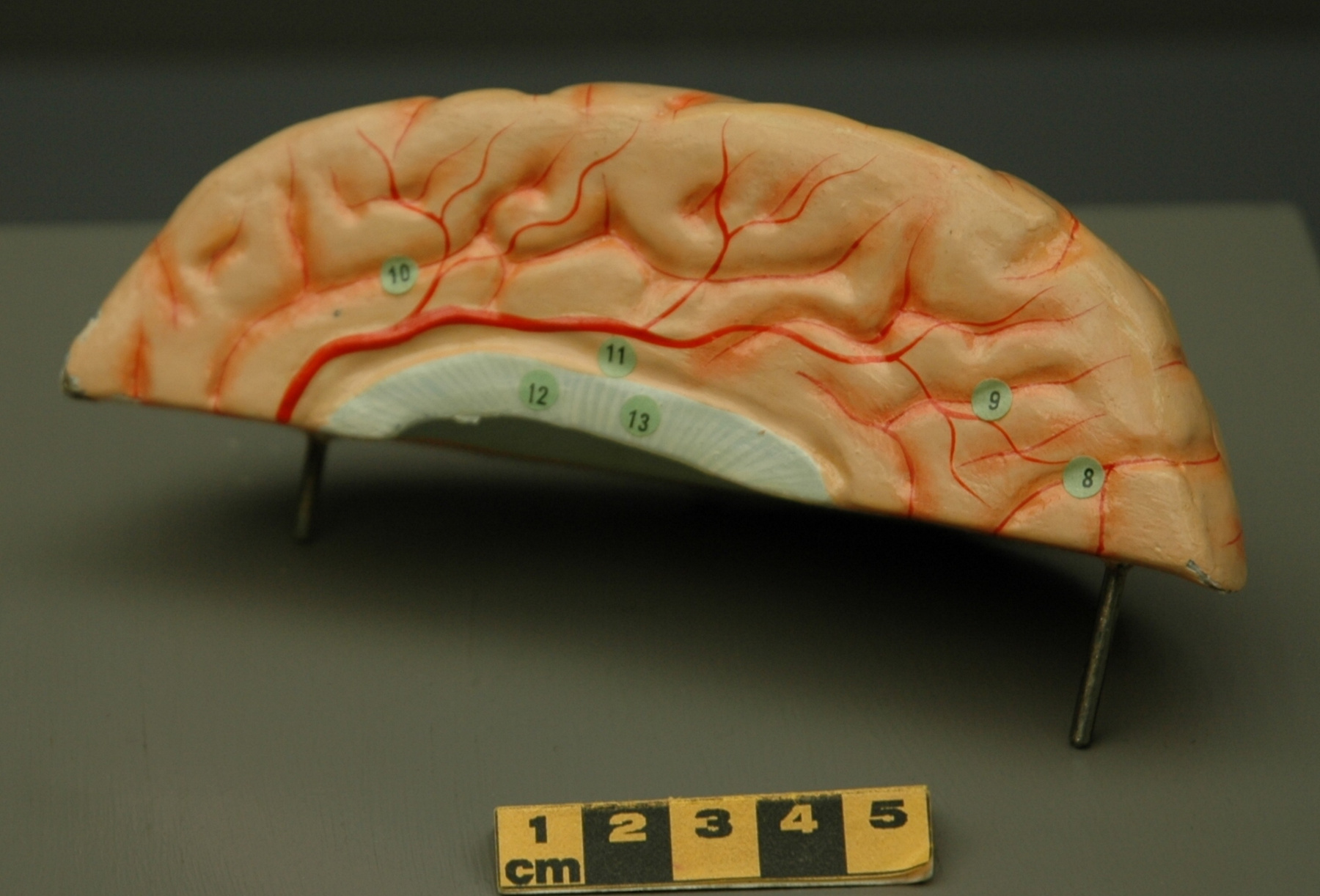
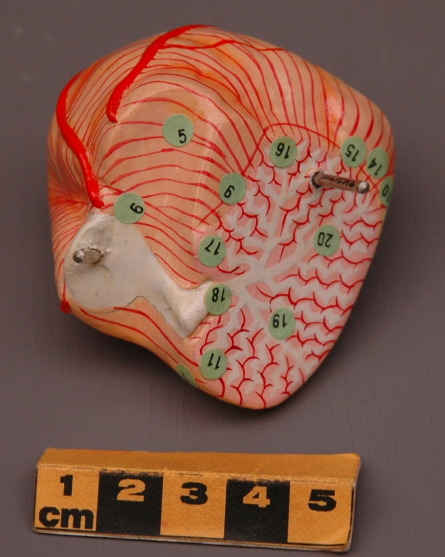
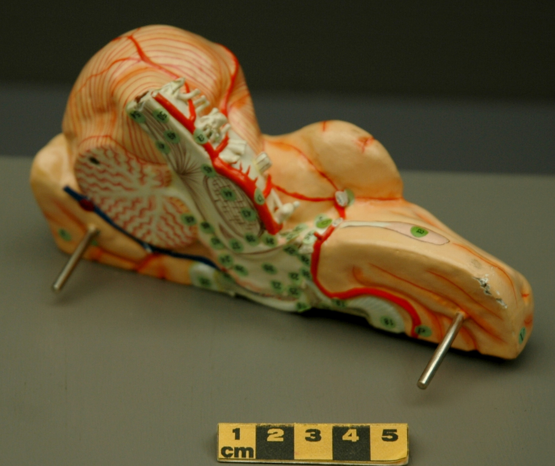
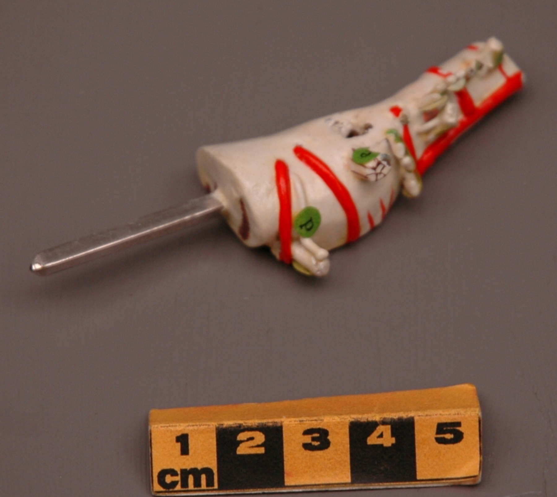
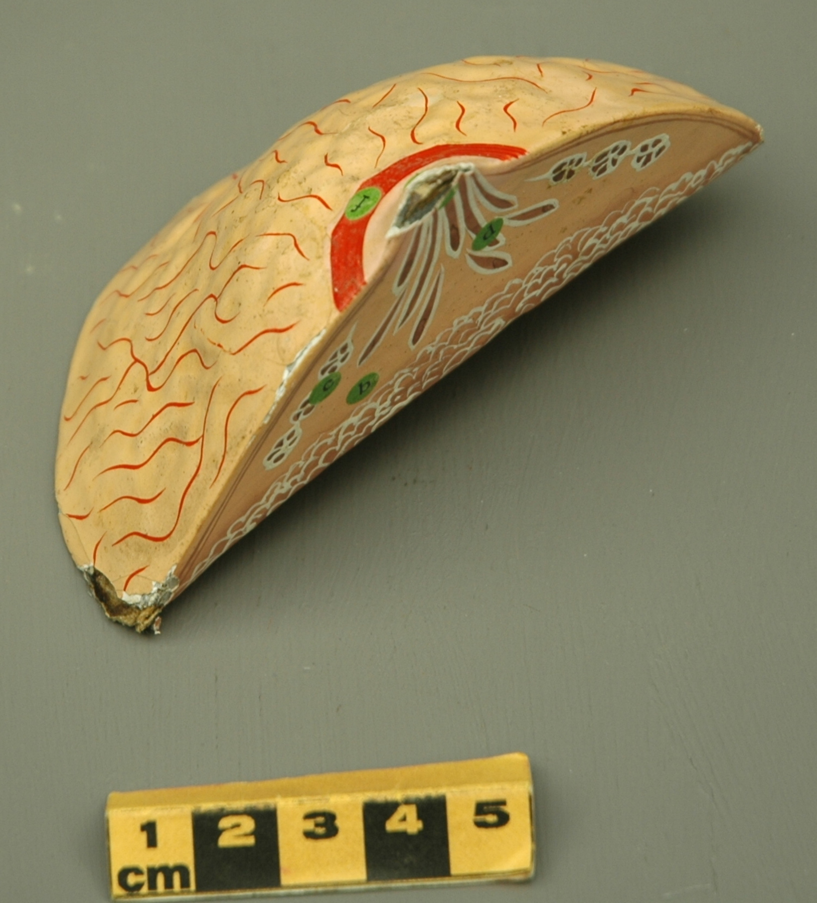
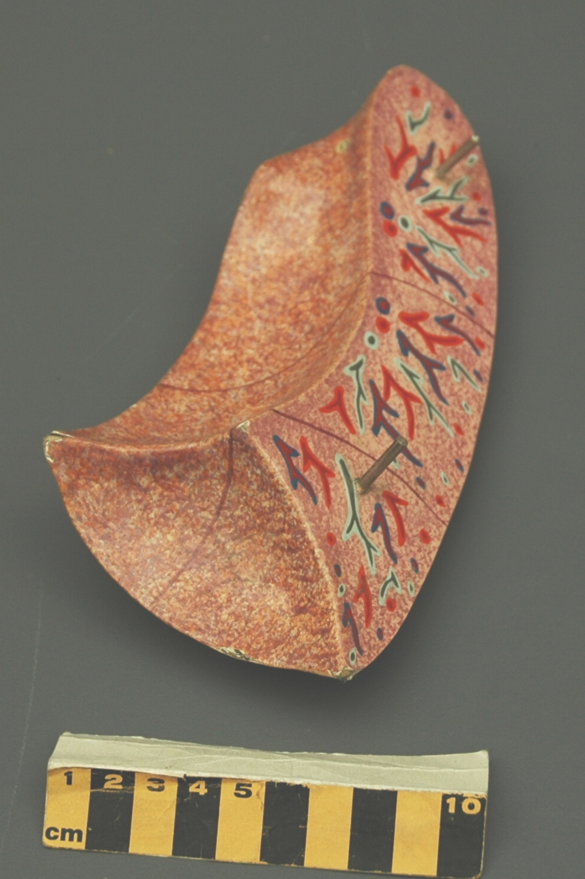
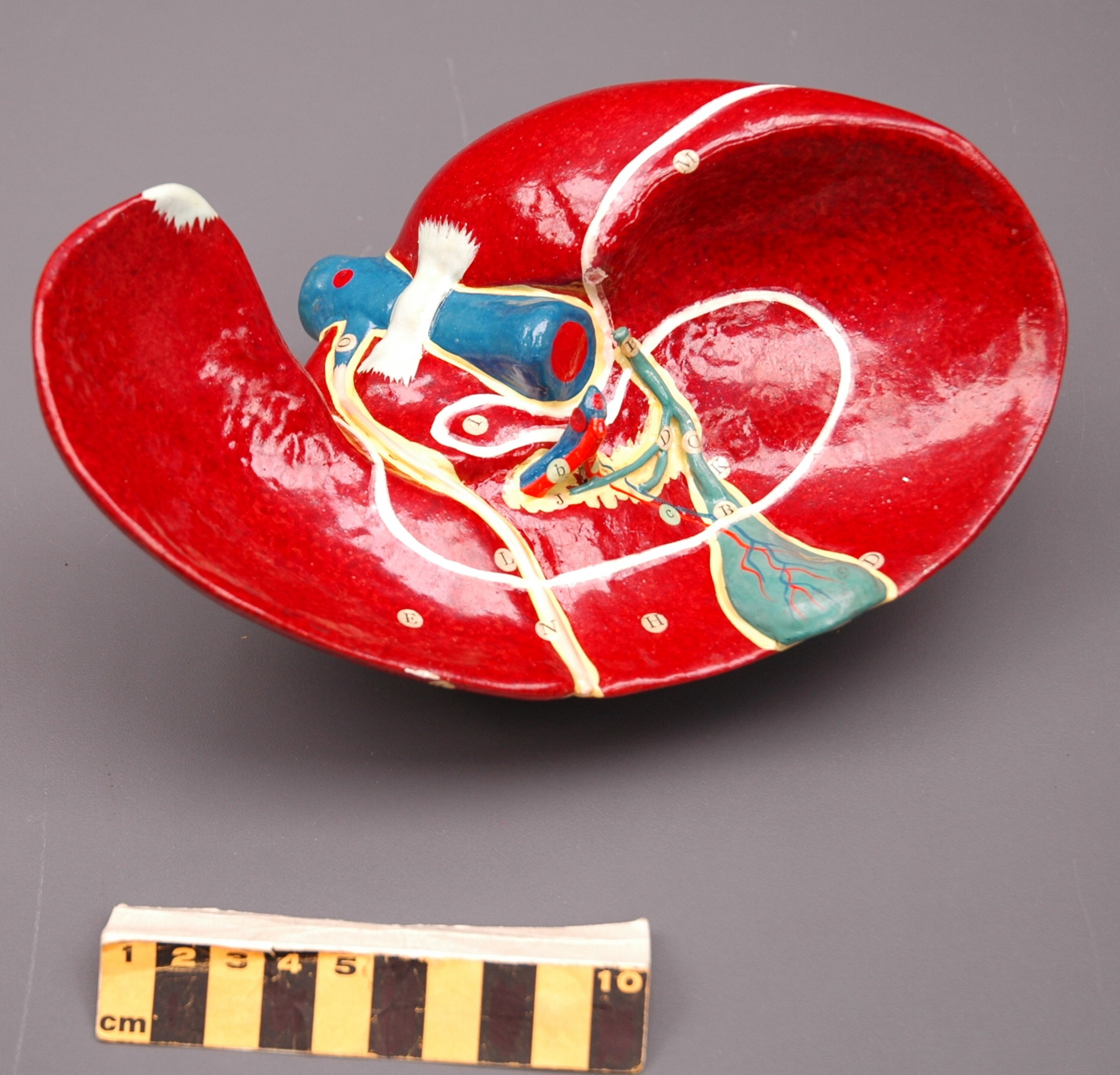
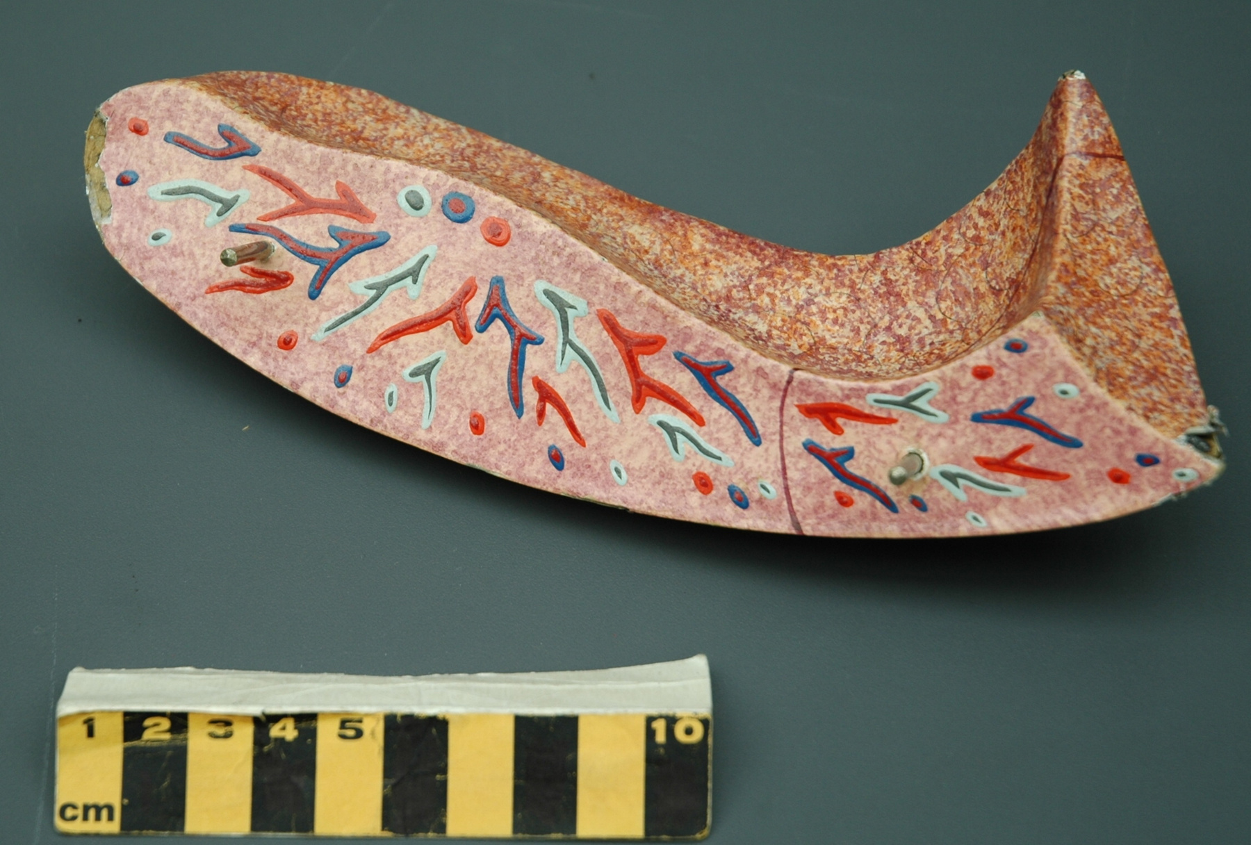
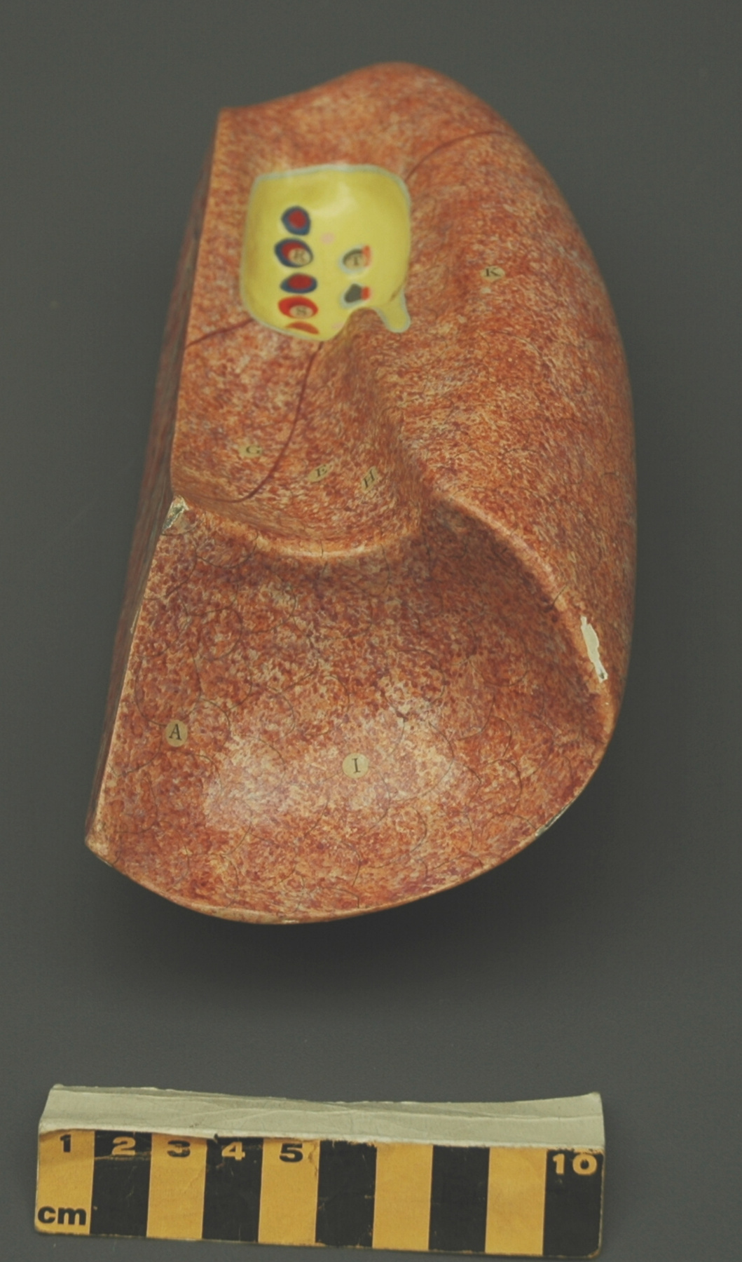
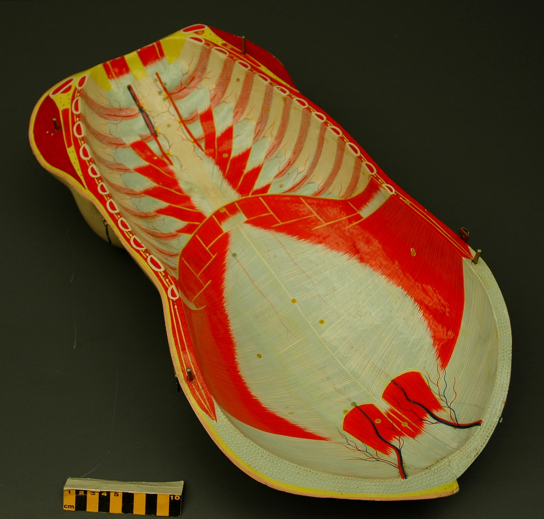
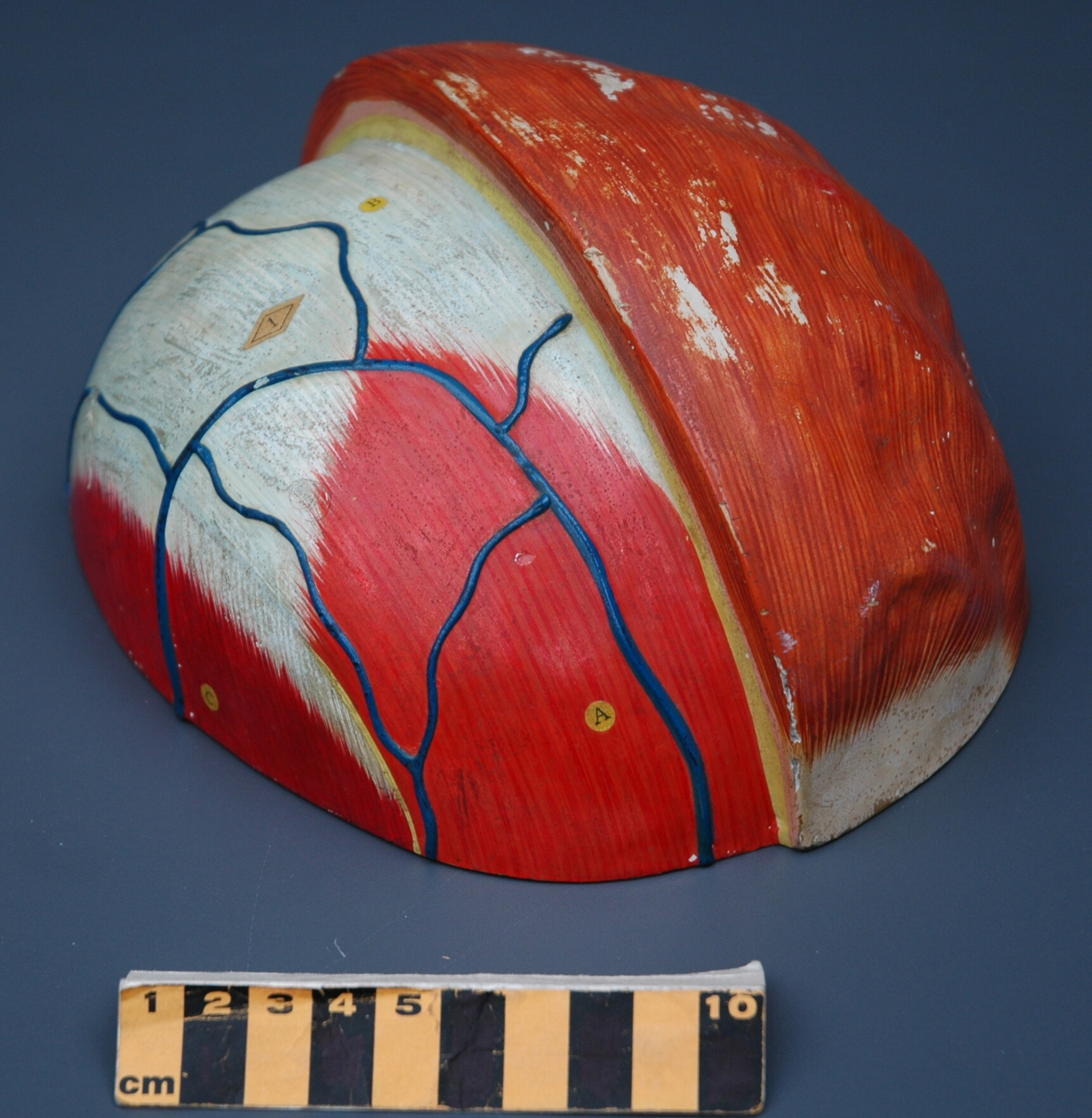
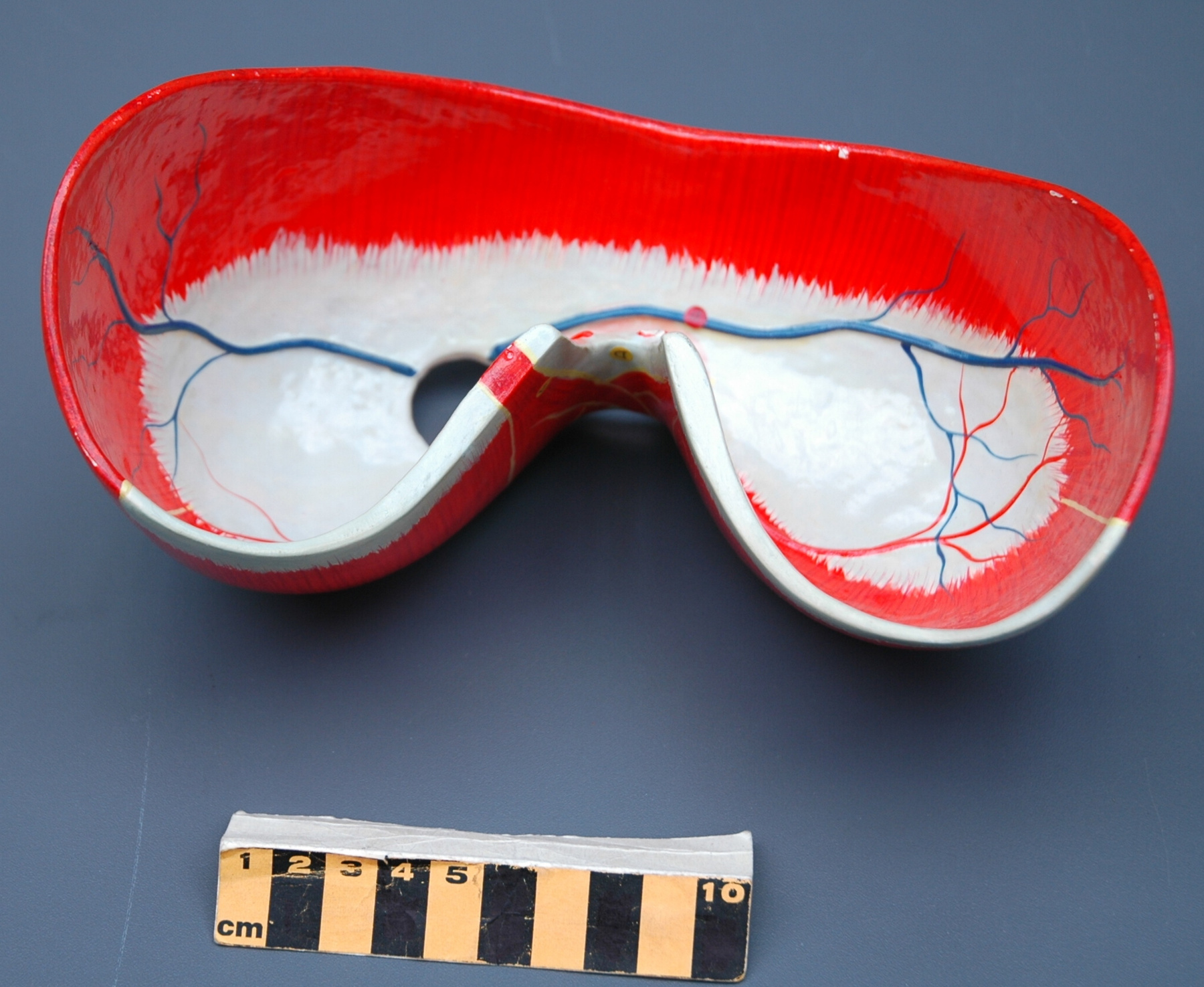
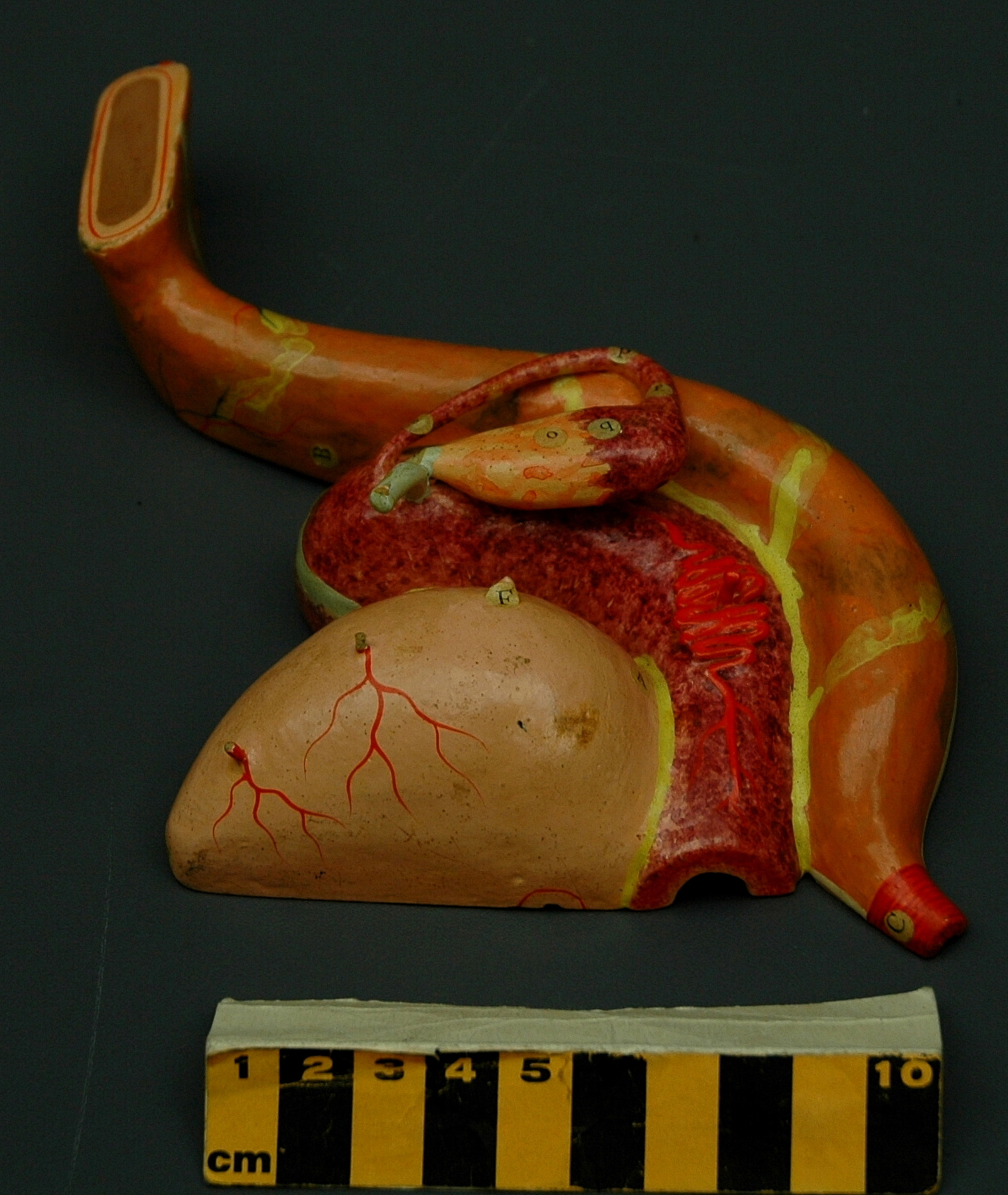
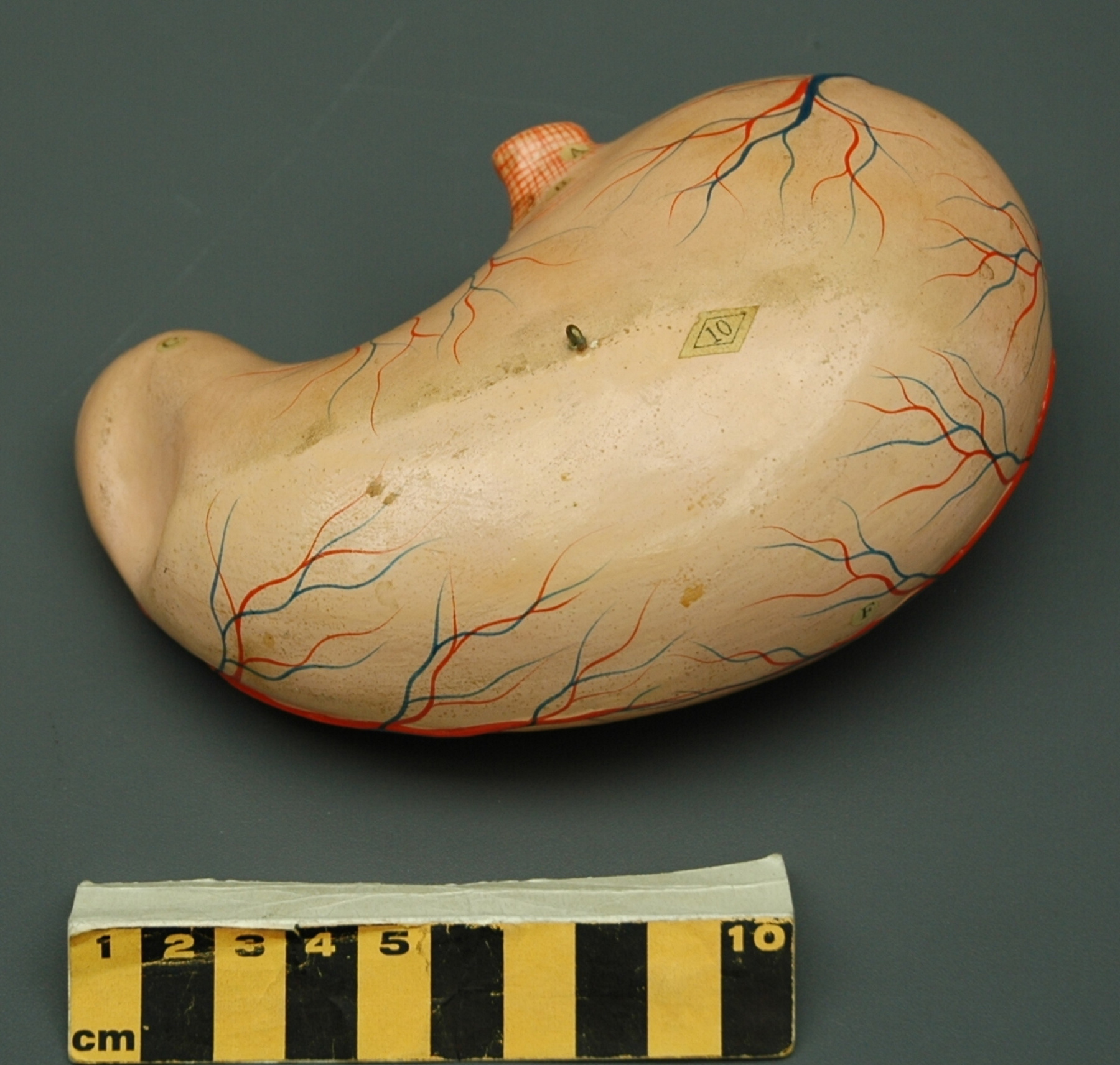
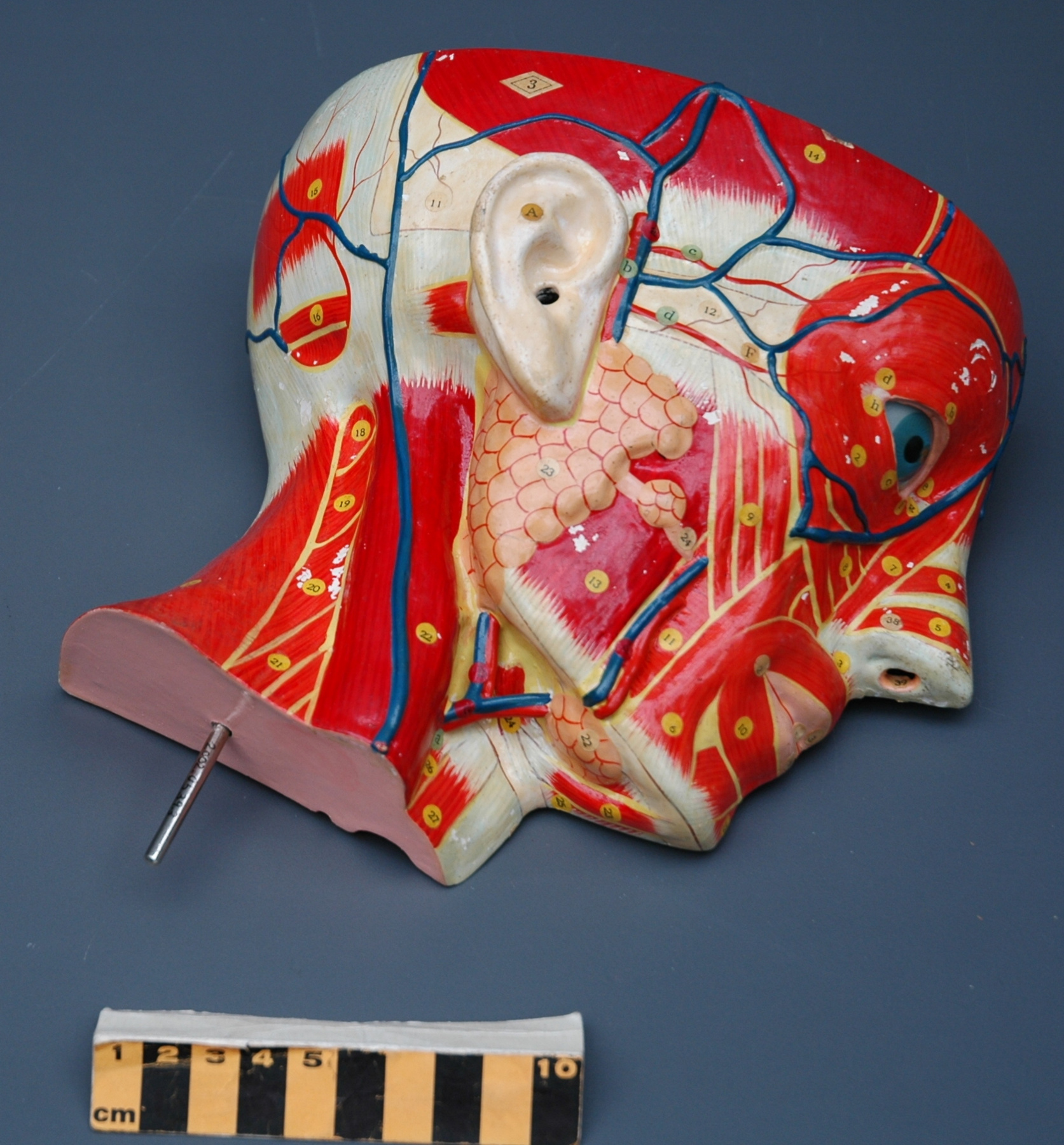
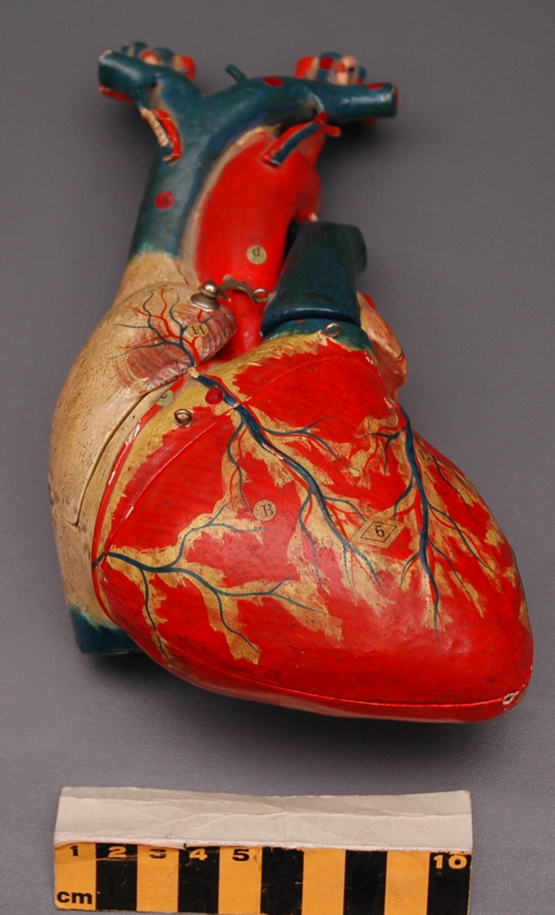
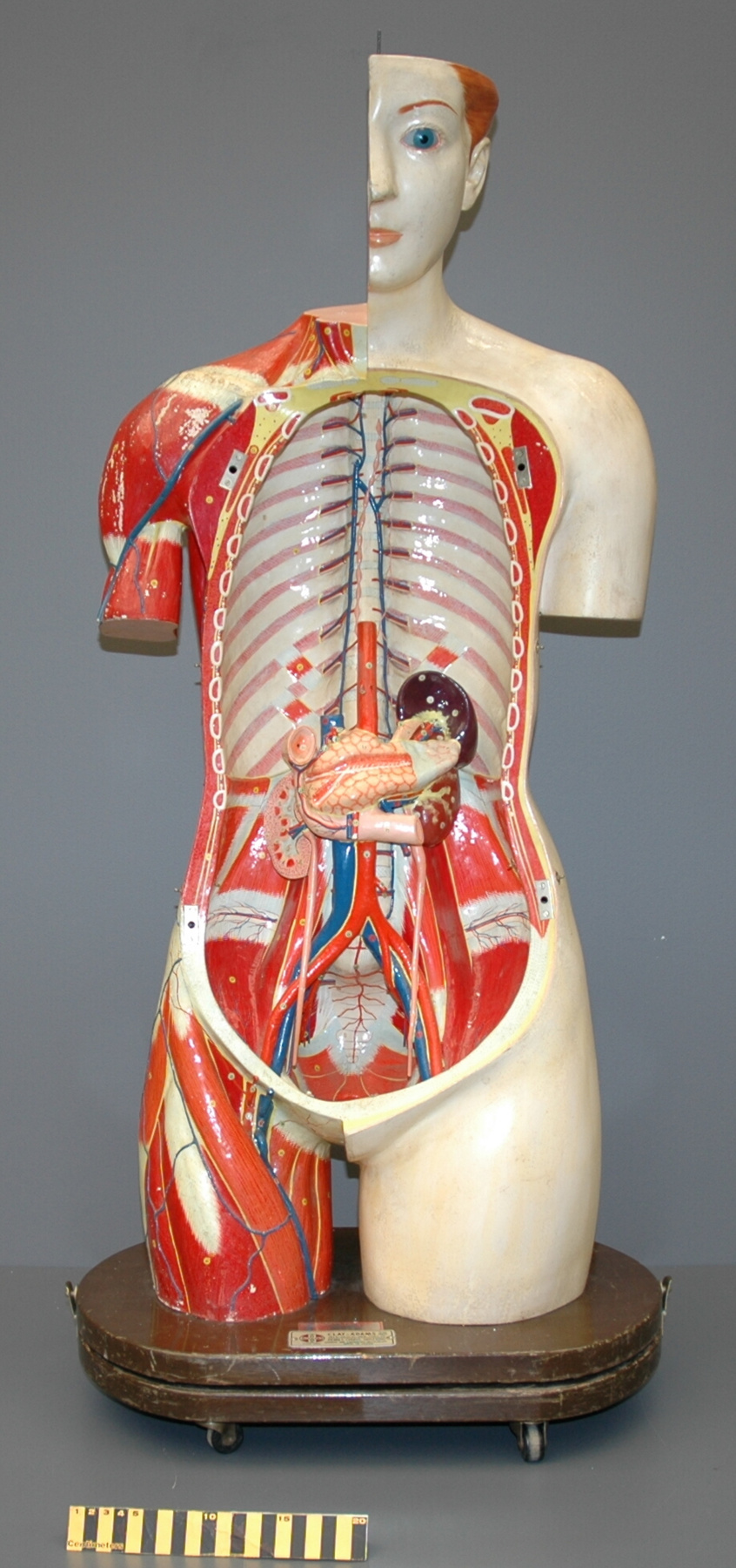
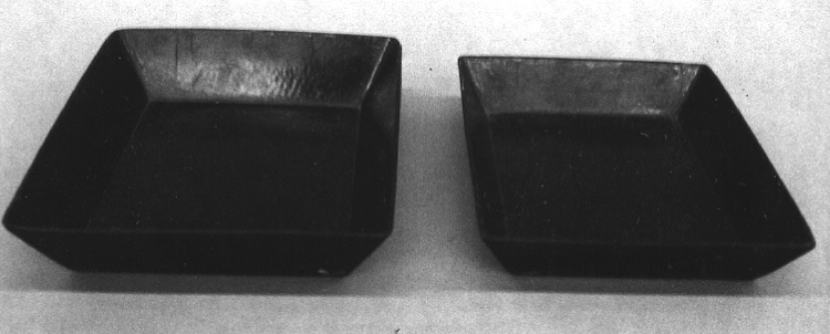
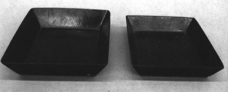
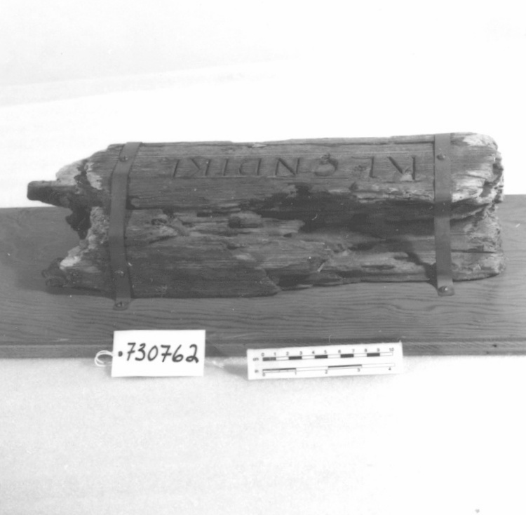
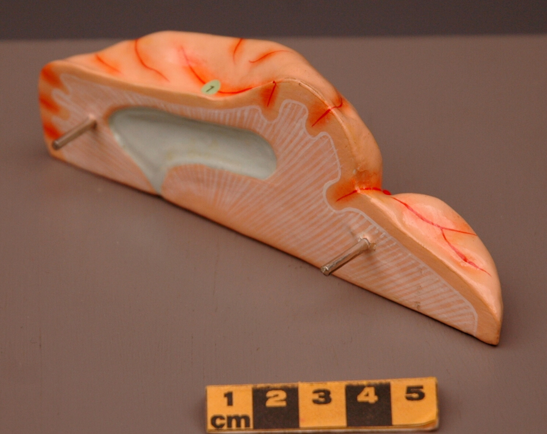
Anatomical model part
Use this image
Can I reuse this image without permission? Yes
Object images on the Ingenium Collection’s portal have the following Creative Commons license:
Copyright Ingenium / CC BY-NC-ND (Attribution-NonCommercial 4.0 International (CC BY-NC 4.0)
ATTRIBUTE THIS IMAGE
Ingenium,
2002.0539.009
Permalink:
Ingenium is releasing this image under the Creative Commons licensing framework, and encourages downloading and reuse for non-commercial purposes. Please acknowledge Ingenium and cite the artifact number.
DOWNLOAD IMAGEPURCHASE THIS IMAGE
This image is free for non-commercial use.
For commercial use, please consult our Reproduction Fees and contact us to purchase the image.
- OBJECT TYPE
- human/torso part/female/life size
- DATE
- 1930–1950
- ARTIFACT NUMBER
- 2002.0539.009
- MANUFACTURER
- Unknown
- MODEL
- Unknown
- LOCATION
- Japan
More Information
General Information
- Serial #
- N/A
- Part Number
- 9
- Total Parts
- 23
- AKA
- portion of brain
- Patents
- N/A
- General Description
- papier-mache on wood; paper labels; silver metal posts.
Dimensions
Note: These reflect the general size for storage and are not necessarily representative of the object's true dimensions.
- Length
- 14.1 cm
- Width
- 4.8 cm
- Height
- 3.0 cm
- Thickness
- N/A
- Weight
- N/A
- Diameter
- N/A
- Volume
- N/A
Lexicon
- Group
- Medical Technology
- Category
- Miscellaneous
- Sub-Category
- N/A
Manufacturer
- AKA
- Unknown
- Country
- Japan
- State/Province
- Unknown
- City
- Unknown
Context
- Country
- Canada
- State/Province
- Ontario
- Period
- This model used c. 1950s+ . Not used after 1980.
- Canada
-
Anatomical model originally owned and used by Dr. Yankoff in his Leaside (east-endToronto) practice, particularly when treating non-English speaking patients. Dr. Yankoff graduated from University of Toronto in 1953, and practiced in Toronto. He died in 1980. - Function
-
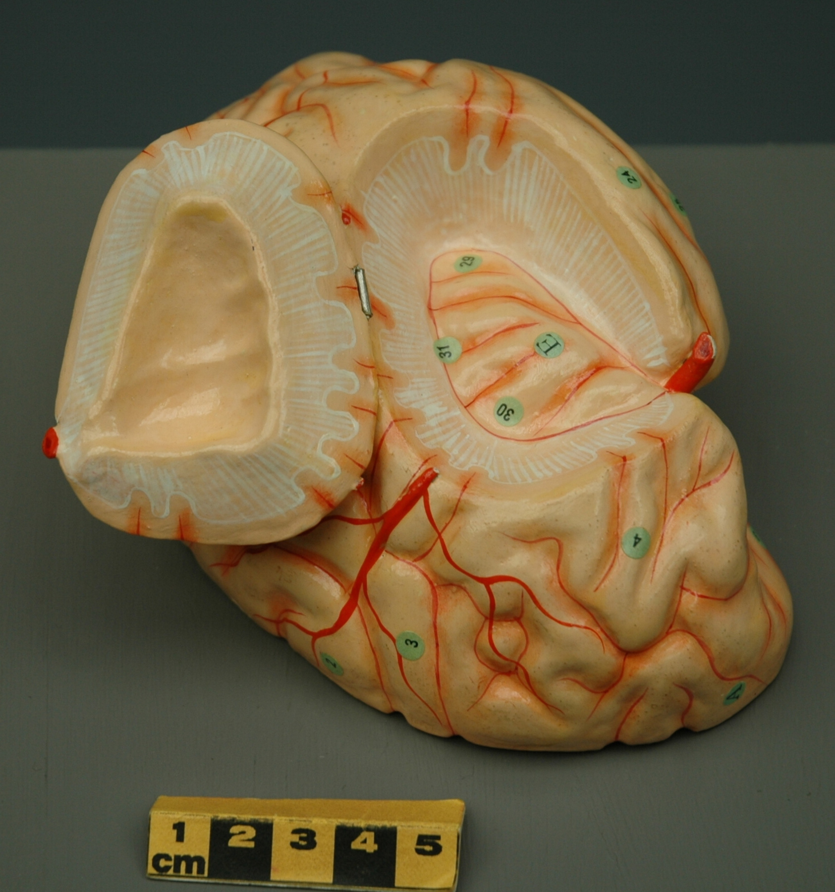
3-D representation of portion of human female brain, possibly including area of temporal lobe. - Technical
-
Anatomical models were popular teaching and demonstration tools, and used in both teaching facilities and practitioner's offices. This model is fairly elaborate, and was originally accompanied by an "atlas" or "key-guide" (now missing) which identified the hundreds of labelled structures. - Area Notes
-
Unknown
Details
- Markings
- None, save decals.
- Missing
- Some small areas of paint/papier mache loss.
- Finish
- This portion of model comprised of papier-mache over wood. Inside & outside surfaces painted: includes red-orange brain and pale blue depression; red, orange and off-white features. Round green paper labels identify selected structures. Two silver metal posts secure this portion within larger brain model mass.
- Decoration
- N/A
CITE THIS OBJECT
If you choose to share our information about this collection object, please cite:
Unknown Manufacturer, Anatomical model part, circa 1930–1950, Artifact no. 2002.0539, Ingenium – Canada’s Museums of Science and Innovation, http://collections.ingeniumcanada.org/en/item/2002.0539.009/
FEEDBACK
Submit a question or comment about this artifact.
More Like This
